
Understanding Pleural Effusion and Drainage
Pleural effusion, often termed “water on the lungs,” involves fluid accumulation between the lungs and chest wall; resuscitation is paramount initially.
Pleural membranes, protecting the lungs, create a pleural space normally containing minimal fluid, but conditions can cause excessive buildup.
Fluid buildup occurs due to disruptions in fluid balance, impacting lung expansion and function, potentially causing chest pain and other symptoms.
Diagnosis relies on clinical data, guiding treatment like thoracentesis to remove fluid, aiding lung function, as detailed in relevant journal articles.
What is Pleural Effusion?
Pleural effusion is characterized by an abnormal accumulation of fluid within the pleural space – the area between the lungs and the chest wall. Commonly referred to as “water on the lungs,” this condition isn’t a disease itself, but rather a sign of an underlying medical issue. The pleura, two thin membranes, normally contain a small amount of lubricating fluid, enabling smooth lung movement during breathing.
However, when this fluid balance is disrupted, excess fluid accumulates, compressing the lung and potentially causing shortness of breath, chest pain, and coughing. The amount of fluid can vary significantly, ranging from small, clinically insignificant amounts to large volumes that severely impair respiratory function. Identifying the cause of the effusion is crucial, as it can stem from heart failure, pneumonia, cancer, kidney disease, or other conditions. Early diagnosis and appropriate management, potentially including drainage procedures, are essential for optimal patient outcomes.
Causes of Fluid Buildup Around the Lungs
Fluid buildup around the lungs, leading to pleural effusion, arises from a variety of underlying conditions impacting the delicate balance of fluid within the pleural space. Heart failure is a common culprit, causing fluid to back up into the lungs. Pneumonia and other infections can trigger inflammation and fluid leakage.
Cancer, both lung and metastatic, frequently causes effusions. Other potential causes include kidney disease, liver cirrhosis, pulmonary embolism, and autoimmune disorders. Sometimes, the cause remains unclear, termed idiopathic pleural effusion. Disruptions in hydrostatic or oncotic pressure, or impaired lymphatic drainage, contribute to fluid accumulation. Accurate diagnosis of the underlying cause is paramount for effective treatment and management of the effusion, often requiring a comprehensive clinical evaluation.
Pleural Membranes and the Pleural Space
Pleural membranes, also known as the pleura, are two thin layers of tissue enveloping each lung. The visceral pleura adheres directly to the lung surface, while the parietal pleura lines the inner chest wall. These membranes create a sealed pleural space between them, normally containing only a few teaspoons of fluid acting as a lubricant during breathing.
This fluid minimizes friction as the lungs expand and contract. The pleural space maintains a negative pressure, crucial for lung inflation. When disrupted, fluid accumulates, leading to pleural effusion. Understanding the structure and function of these membranes is vital for comprehending how fluid buildup impacts respiratory mechanics and necessitates interventions like drainage procedures to restore normal lung function and alleviate symptoms.

Pleurx Drainage: An Overview
Pleurx drainage offers a method for managing pleural effusion, differing from traditional thoracentesis; it’s indicated for recurrent fluid buildup and improved comfort.
What is Pleurx Drainage?
Pleurx drainage represents a specialized technique for managing recurrent pleural effusions, often stemming from conditions like cancer or heart failure. Unlike standard thoracentesis, which is a one-time fluid removal, Pleurx utilizes a small, indwelling catheter placed in the pleural space.
This catheter allows patients – or their caregivers – to drain fluid at home, on a scheduled basis, improving quality of life and reducing the need for frequent hospital visits. The procedure aims to alleviate symptoms like shortness of breath and chest discomfort caused by fluid accumulation.
It’s a less invasive alternative to more complex procedures like pleurodesis, offering a comfortable and convenient method for ongoing fluid management. The system facilitates patient empowerment and self-care in managing their condition.
Pleurx vs. Traditional Thoracentesis
Traditional thoracentesis is a single-event procedure, involving a needle insertion to remove pleural fluid for diagnosis or temporary relief. Conversely, Pleurx drainage employs a small, tunnelled catheter left in situ, enabling repeated, intermittent fluid drainage over an extended period.
Thoracentesis typically occurs in a hospital setting, requiring healthcare professionals for each removal. Pleurx, however, empowers patients to manage drainage at home, reducing hospital visits and improving convenience. While thoracentesis provides immediate relief, Pleurx offers sustained management of recurrent effusions.
Pleurx is particularly beneficial for patients needing frequent drainage, avoiding the discomfort and logistical challenges of repeated needle punctures. It’s a shift towards proactive, patient-centered care, offering a more sustainable solution for chronic fluid buildup.
Indications for Pleurx Drainage
Pleurx drainage is primarily indicated for patients experiencing recurrent, symptomatic pleural effusions – fluid buildup around the lungs – that necessitate repeated drainage. This is especially relevant for individuals with conditions like malignancy, heart failure, or complicated pneumonia, where fluid reaccumulates frequently.
Patients requiring frequent thoracentesis, experiencing discomfort with repeated needle punctures, or facing logistical difficulties accessing healthcare facilities are strong candidates. Pleurx offers a convenient, patient-managed alternative, reducing hospital visits.
Eligibility extends to those who can reliably manage the catheter and drainage process at home, adhering to proper hygiene and monitoring protocols. It’s a suitable option when prolonged, intermittent drainage is anticipated, improving quality of life and minimizing healthcare burden.

Pleurx Drainage Procedure: Preparation
Initial clinical data, guided by diagnosis, informs patient assessment for Pleurx eligibility; gather data and ensure appropriate equipment is readily available;
Initial Clinical Data Gathering
Comprehensive data collection is the foundational step, meticulously guided by the evolving differential diagnosis. This process begins with a thorough patient history, focusing on the onset, duration, and characteristics of symptoms like chest pain and shortness of breath. A detailed review of past medical conditions, including any history of pneumonia, heart failure, or malignancy, is crucial.
Furthermore, a complete medication list, including over-the-counter drugs and supplements, must be obtained. Physical examination findings, such as auscultation for diminished breath sounds or percussion for dullness, contribute significantly. Relevant laboratory investigations, including complete blood count, electrolytes, renal function tests, and coagulation studies, are essential. Prior imaging studies, like chest X-rays or CT scans, should be reviewed to assess the extent and characteristics of the pleural effusion.
This meticulous approach ensures informed decision-making regarding patient suitability for Pleurx drainage.
Patient Assessment and Eligibility
Patient assessment for Pleurx drainage necessitates a careful evaluation of several key criteria. Individuals experiencing symptomatic pleural effusion, unresponsive to conservative management, are primary candidates. Eligibility extends to those with recurrent effusions requiring repeated thoracentesis, potentially benefiting from the convenience of indwelling catheter drainage.
Specifically, patients with complicated pneumonia, as evidenced by studies examining data from 2004-2009, may qualify. However, contraindications include significant coagulopathies, uncorrectable bleeding disorders, and active pulmonary infections requiring immediate intervention.
A thorough assessment of the pleural space via imaging is vital to rule out loculations or trapped lung. Furthermore, the patient’s overall functional status and ability to manage the catheter independently, or with caregiver support, are crucial considerations. Careful evaluation ensures optimal outcomes and minimizes potential risks.
Necessary Equipment and Supplies
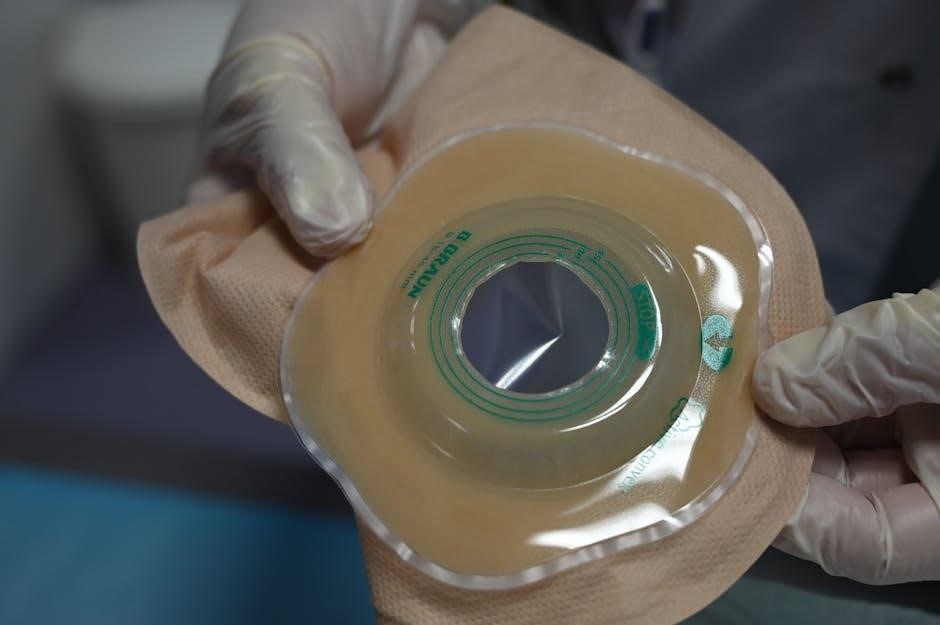
Pleurx drainage requires a meticulously prepared equipment set. Essential items include a dedicated Pleurx catheter of appropriate size, sterile gloves, antiseptic solution (chlorhexidine or povidone-iodine), and sterile drapes to maintain a sterile field.
Local anesthetic (lidocaine) is crucial for pain management during catheter insertion. Additional supplies encompass a scalpel, syringe for local anesthesia administration, guidewire, and a three-way stopcock to facilitate fluid drainage and prevent backflow.
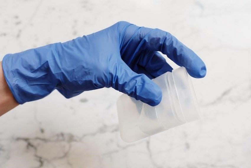
Collection containers for pleural fluid, sterile dressings, and securement devices for the catheter are also necessary. Furthermore, resuscitation equipment, including oxygen and emergency medications, should be readily available. Finally, appropriate personal protective equipment (PPE) for the healthcare provider is paramount for safety.
Pleurx Drainage Procedure: Step-by-Step Instructions
Catheter insertion utilizes a guidewire technique, followed by fluid drainage via a three-way stopcock; monitoring is continuous throughout the process.
Catheter Insertion Technique
Local anesthesia is crucial for patient comfort during catheter placement. Utilizing ultrasound guidance, the intercostal space is identified, ensuring avoidance of vasculature and lung tissue. A small incision is made, followed by blunt dissection to create a tract for the catheter.
A guidewire is then advanced into the pleural space, confirming correct positioning via real-time imaging. The catheter is threaded over the guidewire, and the guidewire is carefully removed.
Secure catheter placement is verified by aspirating pleural fluid. The catheter is then secured to the skin with sutures and a sterile dressing, preparing for the subsequent fluid drainage process. Strict adherence to aseptic technique minimizes infection risk throughout this critical step.
Fluid Drainage Process
Fluid drainage begins once the catheter is securely positioned within the pleural space. Typically, drainage occurs via gravity, utilizing a collection bag positioned below the chest level. The rate of drainage is carefully monitored, avoiding rapid fluid removal which can induce complications like hypotension or pulmonary edema.
Intermittent clamping of the catheter may be employed to assess lung expansion and tolerance to fluid removal. Throughout the process, vital signs – including heart rate, blood pressure, and oxygen saturation – are continuously observed.
The volume of drained fluid is meticulously recorded, providing valuable information regarding the effusion’s characteristics and the patient’s response to treatment. Drainage continues until clinical improvement is noted or a predetermined volume is removed.
Monitoring During Drainage
Continuous monitoring of the patient is crucial throughout the Pleurx drainage procedure. Vital signs – including heart rate, blood pressure, respiratory rate, and oxygen saturation – must be assessed frequently, typically every 15-30 minutes initially, then less often as stability is established.
Lung sounds are auscultated before, during, and after drainage to evaluate for improvement and detect potential complications like pneumothorax. Observe for signs of respiratory distress, such as dyspnea or increased work of breathing.
Drainage volume and characteristics (color, clarity) are meticulously documented. Any sudden changes in drainage rate or appearance should be promptly reported. ECG monitoring may be considered for patients with pre-existing cardiac conditions.

Post-Drainage Care and Instructions
Immediate monitoring assesses vital signs and lung sounds; catheter site care prevents infection, and patients must report any complications promptly.
Follow-up appointments ensure complete recovery, while understanding potential issues is vital for optimal patient outcomes and continued well-being.
Immediate Post-Procedure Monitoring
Following Pleurx drainage, meticulous monitoring is crucial for early detection of potential complications. Vital signs – including heart rate, blood pressure, respiratory rate, and oxygen saturation – should be assessed frequently, typically every 15-30 minutes initially, then extending to hourly intervals as the patient stabilizes.
Lung auscultation is essential to confirm lung expansion and rule out pneumothorax, listening for diminished or absent breath sounds on the treated side. Continuous pulse oximetry provides ongoing assessment of oxygenation. Observe the patient for signs of respiratory distress, such as shortness of breath, increased work of breathing, or changes in mental status.
Pain levels should be regularly evaluated and managed appropriately with prescribed analgesics. The catheter insertion site must be inspected for bleeding, hematoma formation, or signs of infection; Document all observations carefully and promptly report any concerning changes to the physician.
Catheter Site Care
Maintaining catheter site integrity is paramount to prevent infection and ensure successful drainage. Daily assessment of the insertion site is required, observing for redness, swelling, warmth, pain, or purulent discharge – indicators of potential infection.
Strict aseptic technique must be employed during dressing changes, typically performed every 24-48 hours, or more frequently if the dressing becomes soiled or saturated. Gently cleanse the skin around the catheter with an appropriate antiseptic solution, such as chlorhexidine or povidone-iodine, following facility protocols.
Secure the catheter with appropriate tape or a stabilization device to prevent dislodgement. Avoid excessive manipulation of the catheter. Educate the patient (and caregivers) on proper site care techniques and the importance of promptly reporting any signs of complications. Document all care provided and observations made.
Recognizing and Reporting Complications
Prompt identification and reporting of complications following Pleurx drainage are crucial for patient safety. Pneumothorax, indicated by sudden shortness of breath or chest pain, requires immediate medical attention. Infection, signaled by fever, chills, redness, swelling, or purulent drainage at the catheter site, necessitates evaluation and potential antibiotic therapy.
Bleeding, ranging from minor oozing to significant hemorrhage, should be reported immediately. Other potential complications include catheter occlusion, fluid leakage around the insertion site, and rarely, empyema.
Patients and caregivers must be thoroughly educated on these potential issues and instructed to report any concerning symptoms without delay. Healthcare professionals should diligently monitor patients post-procedure and document any adverse events, ensuring timely intervention and optimal outcomes.

Potential Complications of Pleurx Drainage
Pleurx drainage, while generally safe, carries risks including pneumothorax, infection, and bleeding; vigilant monitoring and prompt intervention are essential for patient well-being.
Pneumothorax
Pneumothorax, a potential complication of pleurx drainage, occurs when air enters the pleural space, causing lung collapse. This happens due to accidental puncture of the lung during catheter insertion. Symptoms can range from mild shortness of breath and chest discomfort to severe respiratory distress, depending on the size of the pneumothorax.

Diagnosis typically involves a chest X-ray, which reveals the presence of air outside the lung. Small pneumothoraces may resolve spontaneously with observation and oxygen therapy. However, larger pneumothoraces often require intervention, such as insertion of a chest tube to re-expand the lung.
Healthcare professionals must be vigilant in monitoring patients post-drainage for signs of pneumothorax, including sudden onset of shortness of breath, chest pain, or decreased oxygen saturation. Early detection and appropriate management are crucial to prevent serious respiratory compromise.
Infection
Infection represents a significant risk associated with any invasive procedure like pleurx drainage. Bacteria can enter the pleural space during catheter insertion or through the catheter tract itself, leading to empyema – a pus-filled pleural infection. Strict adherence to sterile technique during the procedure is paramount to minimize this risk.
Signs of infection may include fever, chills, redness, swelling, or increased pain at the catheter site, alongside purulent drainage. A pleural fluid analysis, including Gram stain and culture, is essential for diagnosis. Prompt treatment with appropriate antibiotics is crucial to prevent sepsis and further complications.
Prophylactic antibiotics are generally not recommended, but diligent monitoring and immediate intervention upon suspicion of infection are vital components of post-drainage care. Maintaining a clean catheter site and educating patients on recognizing infection symptoms are also key.
Bleeding
Bleeding is a potential, though generally minor, complication of pleurx drainage. The pleura and intercostal vessels are susceptible to injury during catheter insertion. Patients on anticoagulants or with underlying bleeding disorders are at increased risk, requiring careful consideration and potential medication adjustments prior to the procedure.
Minor bleeding at the insertion site is common and usually self-limiting with local pressure. However, more significant bleeding, manifesting as hematoma formation or hemothorax (blood in the pleural space), requires immediate attention. Monitoring vital signs and observing for signs of blood loss are crucial.
Management of significant bleeding may involve catheter removal, application of pressure, and, in rare cases, blood transfusion or surgical intervention. A pre-procedure coagulation profile helps assess bleeding risk and guide appropriate management strategies.

Pleurx Drainage in Pediatric Patients
Children with complicated pneumonia, aged 18 or less, may be eligible for pleural drainage if discharged from hospitals between 2004 and 2009.
Specific considerations for pediatric drainage involve adapting techniques and dosages, prioritizing safety, and addressing unique anatomical and physiological factors.
Eligibility Criteria for Children
Determining eligibility for Pleurx drainage in pediatric patients requires careful assessment, focusing on children diagnosed with complicated pneumonia. Specifically, the research indicates that patients 18 years of age or younger, who received a pleural drainage procedure due to this condition, were included in a study examining outcomes.
Hospital discharge dates played a crucial role in study inclusion, with eligible patients being those discharged from participating hospitals between January 1, 2004, and June 30, 2009. This timeframe defines a specific cohort for analysis.
Beyond these dates, further criteria likely involve evaluating the volume and characteristics of the pleural effusion, the child’s overall health status, and the presence of any contraindications to the procedure. A comprehensive clinical evaluation is essential before proceeding.
Individualized assessment is paramount, considering the child’s age, weight, and underlying medical conditions to ensure the safety and efficacy of Pleurx drainage.
Specific Considerations for Pediatric Drainage
Pediatric Pleurx drainage demands nuanced approaches compared to adult procedures. Children’s smaller anatomical structures necessitate meticulous catheter insertion technique and careful monitoring to minimize complications like pneumothorax or bleeding.
Pain management is crucial, requiring age-appropriate analgesia and distraction techniques to ensure patient comfort throughout the procedure and post-drainage recovery. Parental involvement and reassurance are also vital components of care.
Fluid volume removal should be carefully controlled, considering the child’s fluid status and renal function to prevent hypovolemia or electrolyte imbalances. Frequent assessment of vital signs and oxygen saturation is essential.
Catheter site care requires diligent attention to prevent infection, utilizing appropriate sterile dressings and monitoring for signs of inflammation or discharge. Close follow-up is necessary to assess for any delayed complications.
Emerging Trends in Pleural Drainage Systems
Automatic pleural drainage systems, like those from Pleural Dynamics, offer alternatives to frequent outpatient visits and invasive procedures for fluid management.
Market analysis indicates growth in chest drainage systems, driven by innovations and increasing demand for less invasive, patient-centered pleural effusion treatments.
Automatic Pleural Drainage Systems
Automatic pleural drainage systems represent a significant advancement in managing recurrent pleural effusions, moving away from frequent manual thoracentesis or prolonged hospital stays for pleurodesis. These systems, such as those developed by Pleural Dynamics, utilize a self-regulating mechanism to continuously drain and manage fluid buildup in the pleural space.
Unlike traditional methods, these systems aim to provide patients with greater autonomy and improved quality of life. They typically involve an indwelling catheter connected to a portable drainage unit that automatically adjusts to maintain negative pressure, facilitating consistent fluid removal. This approach minimizes the need for frequent medical interventions and reduces the burden on healthcare resources.
The development and adoption of these systems are driven by a desire for more effective and patient-friendly solutions for managing chronic pleural effusions, particularly in cases where repeated fluid accumulation significantly impacts a patient’s well-being.
Chest Drainage System Market Analysis
The chest drainage system market is experiencing substantial growth, driven by the increasing prevalence of pleural effusions and advancements in drainage technologies. Analysis, spanning from 2023 to 2033, indicates a rising demand for both pleural drainage systems and specialized pleural drainage system kits.
Key factors fueling this expansion include a growing geriatric population, rising incidence of cancer (a common cause of pleural effusion), and the adoption of minimally invasive procedures like Pleurx drainage. Manufacturers are focusing on developing innovative systems offering improved efficiency, patient comfort, and reduced complication rates.
The market is segmented by product type, end-user (hospitals, clinics, etc.), and geography. North America currently holds a significant market share, but Asia-Pacific is projected to exhibit the fastest growth due to increasing healthcare expenditure and improving access to medical care.

Resources and Further Information
Formal guidelines and evidence-based practices are crucial; consult journal articles for detailed insights into pleural drainage, including Pleurx procedures.
Accessing research provides current understanding of optimal techniques and patient management, enhancing clinical decision-making and improving outcomes.
Formal Guidelines and Evidence-Based Practices
Current clinical practice regarding pleural effusion and drainage, including Pleurx, is continually evolving, necessitating adherence to established guidelines. A case vignette approach, as seen in medical journals, often highlights common clinical problems and evidence-based solutions.
While a specific, universally adopted “Pleurx drainage instructions PDF” may not exist as a single document, best practices are disseminated through professional societies and published research. These resources emphasize thorough patient assessment, appropriate catheter selection, and meticulous aseptic technique during the procedure.
Post-drainage monitoring protocols, including assessment for pneumothorax and infection, are also critical components of evidence-based care. Staying updated with the latest journal articles and research findings ensures clinicians provide optimal and safe Pleurx drainage for their patients, aligning with current standards.
Relevant Journal Articles and Research
Research concerning Pleurx drainage, while growing, isn’t always consolidated into a single “Pleurx drainage instructions PDF.” However, numerous journal articles illuminate best practices. Studies analyzing data from pediatric patients (ages 18 and under) receiving pleural drainage for complicated pneumonia between 2004-2009 offer valuable insights.
Market analysis reports, focusing on chest drainage systems from 2023-2033, detail emerging trends and opportunities for manufacturers, indirectly reflecting advancements in drainage techniques. Furthermore, investigations into automatic pleural drainage systems, like those developed by Pleural Dynamics, showcase innovative approaches.
Accessing databases like PubMed and Google Scholar using keywords like “pleural effusion drainage,” “Pleurx,” and “thoracentesis” yields relevant publications. These resources provide evidence-based guidance on procedure protocols, complication management, and patient selection criteria, informing clinical decision-making.